Nahoko Kasai, Yasuhiko Jimbo, and Keiichi Torimitsu
Materials Science Laboratory
The brain, which consists of neurons and many other different types of cell in a complex network, provides efficient functions including transfer, storage and other forms of processing of a large quantity of information. Numerous attempts have been made to understand these functions, however, few studies have been able to obtain real-time two- or three-dimensional information with precision and high special resolution, which is essential if we are to comprehend how the brain operates.
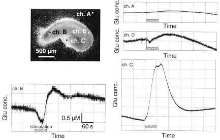
The release of neurotransmitters from neurons and their distribution are also important issues in terms of understanding the mechanisms of memory and learning. However, few studies have succeeded in monitoring the distribution of neurotransmitters themselves. Rather, reports have described the distribution of receptor subunits, which do not inform us of the activity of the receptor and its dynamics. We focus on glutamate (Glu), one of the neurotransmitters in the cortex or hippocampus and have monitored the real-time Glu concentration with a view of imaging and animating its two-dimensional distribution. In this study, we have succeeded in monitoring the Glu concentration at multiple positions in a hippocampal slice simultaneously [1]. We fabricated an electrochemical Glu sensor array by modifying an electrode array (each size: 50X50 μm2) with enzymes and an electron transfer mediator.
We then placed the cultivated slice on the array and measured the Glu concentration
at selected sensors in the array. When we introduced a stimulant into the
external medium, the sensors indicated the different amounts of Glu released
as a result of the receptor stimulation (Fig. 1). This result demonstrates
the diversity of the receptors, their distribution and their dynamics.
Our glutamate sensor array could prove to be a powerful tool for understanding
the role of glutamate in the brain. We will continue our work in order
to realize the imaging of its distribution.
[1] N. Kasai et al., Neurosci. Lett. 304 (2001) 112.

| Fig. 1. | Glutamate concentration profiles at different positions in hippocampus. |