Materials Science Laboratory
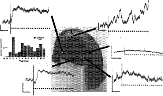
ü@Magnesium ion has been known to play an important role in biochemical processes and development. However, its role in neural processes is still unclear. In this research, we focused on the effect of magnesium ion on the neural activities in rat hippocampal neurons. The neuronal responses, such as electrical bursts and glutamate release were investigated in rat hippocampus under low magnesium conditions. Measurements have been carried for dissociated cell cultures and slice preparations. Electrical bursts were measured using a 64-channel ITO microelectrode array. The size of the electrode was 10-50 ā╩m each side. The array was fabricated with lithographical techniques and directly connected to 64-channel amplifier. Using this array, spatial distribution of the electrical activities could be measured. Transient changes of glutamate release were measured using an enzyme-based electrochemical detection method. The method includes glutamate oxidase, horseradish peroxidase and mediator polymer. They were placed on each microelectrode. Hippocampal neurons including slices were obtained from either P2 or P8 Wistar rat and cultivated them on a culture dish or a porous membrane for 7-14 days.
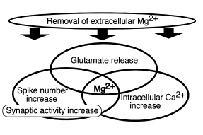
ü@Electrical bursts turned to be highly active in a magnesium-free medium (MGF). A transient increase induced by the MGF was detected in the glutamate concentrations in the CA1, CA3 and dentate gyrus within a few minutes. Intracellular Ca concentration was also increased transiently. The NMDA receptor antagonist, MK801 suppressed the increase only in the CA1, but not in the CA3 and dentate gyrus. The results may indicate the diversity of NMDA receptor distribution and its dynamics in the hippocampus. Magnesium ion could be a key in the control of the neural activities in rat hippocampus.
[1] N. Kasai et al., Neurosci. Lett. 304 (2001) 112.
[2] K. Torimitsu et al., Gordon Res. Conf. 3 (2002)
 |
 |
||||
|
|