Recently, neuron-nanostructure integrated devices have been attracting a lot of interest in relation to understanding signal transfer mechanisms. In such devices, signals from single neurons, e.g., neuron action potentials, are transmitted extracellularly via conductive materials, and hence the interface between single neurons and conductive materials plays an important role. However, there have been few investigations of this interface because of the difficulty involved in direct and high-resolution observation with conventional optical microscopy. We employed a focused ion beam (FIB)/SEM combined system, which enabled the cross-sectional observation of neurons cultivated on the surfaces of conductive materials. We also examined the relationship between the interface structure and neuron-substrate affinity by using fluorescence microscopy.
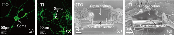
Neurons were prepared from rat cortex and cultivated on two types of conductive thin films: indium tin oxide (ITO) and Ti. The cultivated neurons were immunostained and observed with a fluorescent microscope. For the FIB/SEM experiments, the cultivated neurons were dehydrated, fixed and finally freeze-dried. Figures 1 (a) and (b) are fluorescence images of the neurons on ITO and Ti, respectively. On the ITO surface, somas tend to aggregate and dendrites grow linearly, indicating the low affinity between neurons and ITO. In contrast, on a Ti surface, very little soma aggregation or straight dendrite growth is observed, implying a high affinity between neurons and Ti. Figures 1 (c) and (d) are cross-sectional SEM images of single neurons on ITO and Ti surfaces, respectively. Although the neurons hardly make contact with the ITO surface as indicated by the wide interspace between the neurons and the ITO, they come into close contact with the Ti surface. These results obtained in fluorescence and FIB/SEM experiments indicate that Ti has the higher biocompatibility.
Our approach allowed high-resolution observation of the interface between single neurons and substrates, which will provide important implications for the control of neuron growth, leading to a quantitative analysis of the neuronal signals from neuron-nanostructure integrated devices.