Conductive Silk Film Electrodefor Manipulation and Electrical Stimulation of Adherent Cells
Action potentials generated by ion channels are involved in a wide range of cellular processes. Microelectrode arrays (MEAs) or transistor biosensors have been developed to stimulate cells or measure electrical signals. However, there is a technical issue as regards controlling the location of cells on the electrodes. This is because the cells adhere randomly to the surface, which hampers experimental reproducibility and accuracy. We thus approach this issue by developing a mobile, conductive film-based interface (nano-pallet) that enables us to manipulate and electrically activate specific cells. We fabricated the nano-pallet by using a combination of silk fibroin hydrogel and PEDOT:PSS [1] to improve transparency, biocompatibility, mechanical stiffness, and electrical conductivity.
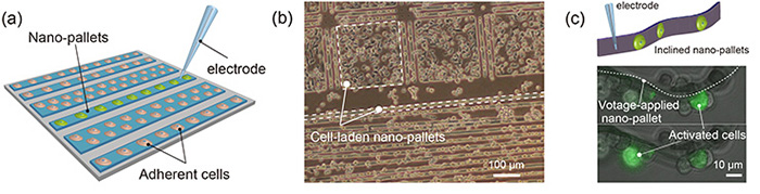
The micropatterned nano-films were formed with a controllable size and shape by using a photolithographic technique [Fig. 1(a)] [2]. The silk fibroin matrix with embedded PEDOT:PSS is optically transparent in both the visible and ultraviolet regions, enabling the observation with various types of microscopes. The FTIR spectra had peaks at 1535 and 1630 cm–1 assigned to the amino-1 band, which indicated that the film contained β-sheet crystalline fractions. The gelled silk fibroin proteins improved the mechanical properties of the film (elastic coefficient = 100 MPa). Interestingly, the fabricated nano-film realized 500 times higher conductivity (at a maximum of 1 mS) than pristine PEDOT:PSS film, implying that the silk fibroin molecules help the rearrangement of the PEDOT:PSS chains and enhance charge transfer in inter-chains or inter-particles.
Since silk fibroin and PEDOT:PSS are highly biocompatible, the cells tend to migrate and proliferate at the nano-pallet surface [Fig. 1(b)]. We micropatterned the film to modulate the behavior of specific cells and activate them while retaining their adhesive property. We further utilized these films to individually measure and apply cellular action potentials to a target cell by incorporating them in capillary electrodes. By applying a voltage to cell-laden nano-films, cells expressing P/Q-type calcium channels (Cav2.1) were selectively activated under a homogeneous condition [Fig. 1(c)]. These properties along with non-cytotoxicity and long-term implantation capability could provide a prototype biocompatible electrode for cells and tissues. We believe that the nano-films can be used for both in vitro electrophysiological analysis and various biomedical applications.
- [1] S. Tsukada et al., PLoS One 7, e33689 (2012).
- [2] T. Teshima et al., Adv. Funct. Mater. 26, 8185 (2016).
|
|
Fig. 1. (a) Schematic illustration of cell-laden nano-pallet electrodes. (b) Micrograph of cell-laden nano-pallets. (c) Merged micrographs of phase-contrast and confocal images of voltage-applied nano-pallets. |