Hidetoshi Nakano, Peixiang Lu, Tadashi Nishikawa, and Naoshi Uesugi
Physical Science Laboratory
The recent development of high-power ultrafast laser technologies has made laser-produced plasmas attractive as potential bright X-ray sources. High-density plasmas created near a solid surface by femtosecond laser pulses emit short X-rays pulses in the energy range from sub-keV to MeV energies. Moreover, these X-rays are synchronized to the incident laser pulse. Such X-rays are extremely important as diagnostic probes in the pump-probe-type experiments for observing the dynamic responses of optically excited materials.
We demonstrated time-resolved measurement of soft X-ray absorption in optically pumped silicon membrane by means of pump-probe spectroscopy and found that soft X-ray absorption in Si near its LII,III edge could be rapidly modulated by an intense laser pulse irradiation [1]. Measurements were carried out using a soft X-ray pulse emitted from a femtosecond laser plasma created on a Ta film as a probe and a 100-fs laser pulse with a wavelength of 790 nm as a pump. The duration of the probe pulse near a photon energy of 100 eV was measured to be 40 ps by using an X-ray streak camera. The sample was 100-nm-thick silicon without supporting structures. When the intensity of the pumping laser pulse on Si membrane was 1010 W/cm2, which is well below the damage threshold, a 5% increase in soft X-ray absorption near LII,III absorption edge was observed as shown in Fig. 1. Figure 1 shows the result when the pump and probe pulses arrived on a sample simultaneously. The most significant dip in the differential transmission spectrum (C) appeared near 99.5 eV. The sharp change in soft X-ray transmission only appeared near 99.5 eV, which is slightly lower than the LII,III edge. This change was observed when the relative time delay was within 40 ps. Therefore, in this case, the dependence of the transmission change at 99.5 eV on the time delay of the probe soft X-ray pulse well fitted the probe pulse shape, as shown in Fig. 2. The origin of this absorption change is most likely an additional absorption line and/or band gap shift due to high-density photo-induced electron-hole plasma creation.
[1] H. Nakano et al., Appl. Phys. Lett. 75 (1999) 2350.
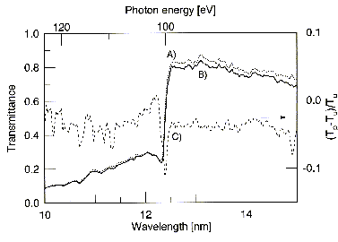
Fig. 1. Transmission spectra near the LII,III edge of Si. Dotted (A) and thick solid (B) curves represent transmission spectra observed with and without laser irradiation. The thin dotted curve (C) shows the differential transmission.
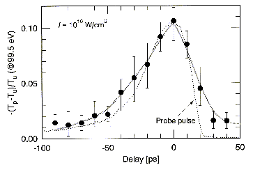
Fig. 2. Depth of the dip in differential transmission at 99.5 eV as a function of the probe soft X-ray pulse delay. The thin dotted curve shows the probe pulse shape measured by an X-ray streak camera.
Back