Å@
Tobias Nyberg, Akiyoshi Shimada, Nahoko Kasai, and Keiich Torimitsu
Materials Science Laboratory
Å@
Å@We have examined the stimulation and recording properties of conjugated
polymer microelectrode arrays as interfaces with neural networks of dissociated
cortical neurons.
Å@The polymer electrodes were electrochemically polymerized from a blend
of poly (3,4-ethylenedioxythiophene)-poly (styrenesulfonate) (PEDOT-PSS)
and ethylenedioxythiophene (EDOT) onto indium tin oxide (ITO) microelectrodes.
Conducting polymers have been utilized to increase the surface roughness
and improve the performance of planar electrodes [1].
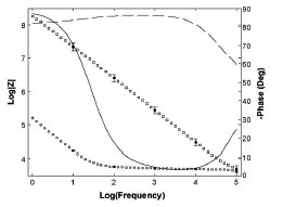
Å@The stimulation properties were investigated as a means of supplying
a neural network with information. The impedance of the polymer electrodes
(circles) was markedly lower than that of the ITO electrodes (squares)
for low and medium frequencies and the phase of the polymer electrodes
(solid line) was lower than that of the ITO electrodes (dashed line) for
medium frequency as shown in Fig. 1. The peak current was proportional
to the applied voltage pulses for polymer electrodes.
Å@Dissociated cortical neurons from Wister rat embryos (embryonic day 18)
were then plated on the electrodes and cultivated to form neural networks.
Spontaneous activity was detected by both bare ITO and polymer electrode
after 5 days in vitro and the bursting frequency increased as the networks
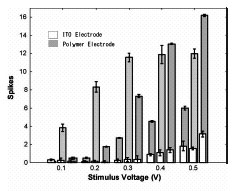
matured. The stimulation efficiency at low voltages was evaluated and referenced
to ITO electrodes. Polymer electrode stimulation evoked a much greater
response from the network than stimulation from ITO electrodes as seen
in Fig. 2. Polymer electrodes could be used at low stimulating potentials
for the efficient stimulation of neuronal tissue for more than one month
and interfacing could be maintained for several months. These results show
that conducting polymer electrodes have the biocompatibility needed microelectrodes
for interfacing with neural networks.
[1] T. Nyberg, et al., J. Neurosci. Methods. 160 (2007) 16.
 |
 |
|||||
|
|