1Materials Science Laboratory, 2Photonics Laboratories, and 3Access Network Service System Laboratories
We have developed a new method for delivering water-soluble chemicals,
broadband light and electrical stimulation using one optical fiber. This
constitutes a new tool in photorelease technology for controlling cellular
chemistry and physiology [1].
First we injected caged glutamate (4-Methoxy-7-nitroindolinyl-caged-L-glutamate)
into a culture consisting of dissociated cortical neurons of an embryonic
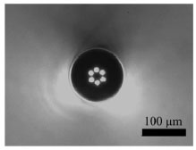
day 18 rat using a holeassisted optical fiber (HAF)[2]. The HAF has a Ge-doped
silica core and six equally spaced holes with a diameter of 14 µm
in the cladding surrounding the core (Fig. 1). The end of the HAF was located
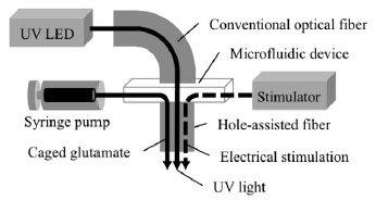
just above the neurons. 250 µM of caged glutamate was injected into
these holes with a syringe pump via a microfluidic device (Fig. 2). To
photorelease caged glutamate, 365 nm ultraviolet (UV) light from an LED
was coupled into the HAF through the microfluidic device. The cortical
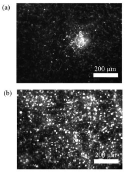
neurons were preloaded with the Ca2+ sensitive fluorescent dye, fluo-4. When the UV light released the glutamate, we observed an increase in the intracellular Ca2+ concentration using fluorescent microscopy [Fig. 3(a)]. We then applied
electrical stimulation from this HAF by utilizing these holes filled with
electrolyte solution and observed an increase in the intracellular Ca2+ concentration [Fig. 3(b)].
The HAF has an outer diameter of 125 µm and an extremely low bending
loss characteristic compared with conventional optical fiber. This HAF
provides the possibility of achieving a less invasive approach to surgery.
[1] A. Shimada et al., Neuroscience2011, 204.27, Washington DC, 2011.
[2] K. Nakajima et al., Photonics Technology Letters, IEEE 15 (2003) 1737.
 |
 |
 |
||||||||
|
|
|