Materials Science Laboratory, *NTT Microsystem Integration Laboratories
Graphene oxide (GO) is an analogue of an oxidized form
of graphene, which contains a number of C-O bonds generated by the oxidization
of C=C double bonds. GO holds an atomically thin sheet-like structure like
graphene, but at the same time it shows many different features from graphene.
For instance GO is an insulator, water-dispersive material and efficient
fluorescence quencher. We designed and built the label-free protein recognition
system on the GO surface using these unique properties [1].
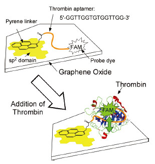
We used GO fixed on a solid surface. The GO surface was
modified using pyrene linker to sp2 domains left in GO and the thrombin
aptamer with a probe dye (FAM) bonded at the other terminus (Fig. 1, top).
Here the aptamer is a single strand DNA that forms a complex with a specific
target molecule. In this study, we used the aptamer for thrombin, an important
protein for blood clotting, to demonstrate the idea.
The recognition mechanism is schematically shown in Fig.
1. Fluorescence from the dye is quenched by the GO at the initial stage
when the modified molecules adsorbed on the GO surface with the strong
interaction between single strand DNA and GO. The fluorescence recovers
when the aptamer recognizes and forms a complex with thrombin (Fig. 1,
bottom). This is because the dye is separated from the GO surface by the
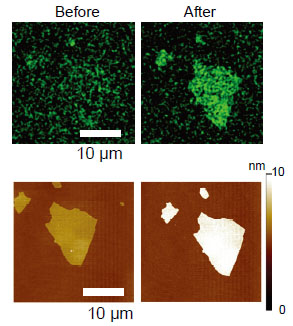
recognition. The validity of the system was confirmed using a single piece
of GO. Fluorescence microscope observations showed that the fluorescence
recovers only on the GO surface after the thrombin addition (Fig. 2, top).
The thickness of GO was increased about 2.9 nm upon the thrombin recognition,
which was traced by atomic force microscope observations for the same GO
piece (Fig. 2, bottom). The experiments demonstrated the successful label-free
detection of thrombin. Our system is advantageous for the operation in
microfluidics because the GO piece is firmly fixed on the solid surface.
With an increase variety of detectable proteins by choosing different aptamers,
it provides a common platform of GO aptasensors.
[1] K. Furukawa et al., J. Mater. Chem. B 1 (2013) 1119.
 |
 |
|||||
|
|