Å@
Koji Sumitomo1, Mime Kobayashi1, Hiroshi Nakashima1, C. S. Ramanujan2, Keiichi Torimitsu1, and J. F. Ryan2
1Materials Science Laboratory, 2University of Oxford
Å@To make a novel bio-nanodevice, uniting nanotechnology with biotechnology, it is vital to investigate individual biomolecular structures and dynamics at molecular-scale in physiological conditions. Only then can we hope to understand how to control the functions of such a device. Atomic force microscopy (AFM) is one of the most powerful tools to observe the biomolecules in their native liquid environment. In this study, we successfully visualized the static crystalline structures of membrane proteins and the dynamic molecular motion of DNA by liquid-state AFM measurements.
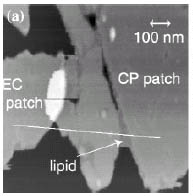
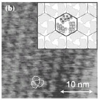
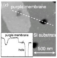
Å@Fig. 1(a) shows topographic AFM image of purple membrane patches on mica. We can distinguish either extracellular (EC) side or cytoplasmic (CP) side of purple membrane from their surface morphology such as the smoothness and from the mechanical characteristics like the stiffness. High-resolution AFM imaging clearly exhibits the 2D crystal of bacteriorhodopsin (bR) protein in the purple membrane, as shown in Fig.1 (b). Recently, we have succeeded in imaging the suspended membrane over artificially fabricated, nano-scale cells on a silicon substrate (Fig. 1(c)). In its native cell membrane, the bR proteins functions as a channel that opens by irradiation of light and pumps protons out of the cell through the membrane. This proton gating function, working on the nano-scale cell, has promising applications in optoelectronic nanodevices [1]. We are now exploring the detailed correlation between the conformational change and optical function of bR on the cell by AFM.
Å@On the other hand, by using a high-speed AFM that is capable of catching video-rate images, we have succeeded in observing biotinylated DNA binding to/dissociating from a streptavidin protein [2]. High-speed imaging enables us to visualize the short DNA strands in motion. We successfully captured real-time images of a biotin of the DNA end binding to a streptavidin. The results indicate that high-speed scanning of AFM is potentially available for the observation of biomolecular dynamic events such as chemical reaction or response to external stimuli in liquid.
Å@Based on the combination of the static and dynamic analysis by AFM with electrochemical approach, we are aiming to design and fabrication novel bio-nanodevices.
[1] K. Sumitomo et al., NTT Technical Review (in press).
[2] M. Kobayashi et al., Ultramicroscopy (in press).
 |
 |
 |
||||||
|
|
|