Materials Science Laboratory
Lipid bilayer, the fundamental component of cell membranes, can form
at the solid-liquid interface by self-organization. We have developed a
new type of microchannel device using this self-spreading characteristic
of the lipid bilayer [1]. The validity of the device has been confirmed
by observing fluorescence resonance energy transfer (FRET). This report
is of the successful application of the device to the quantitative determination
of FRET efficiency [2].
L-α-PC (extracted from egg yolk) containing 1 mol % of NBD- (a dye
for donor) or Texas Red (TR, a dye for acceptor)-conjugated lipid is prepared.
They are self-spread from the each side of the straight line pattern with
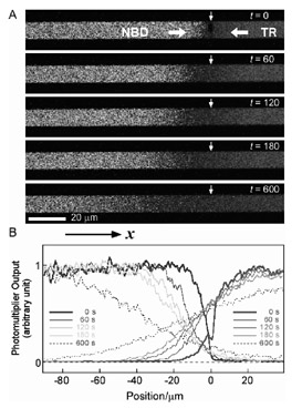
a 10 mm width. Figure 1A shows the confocal laser scanning microscope images
from the moment the lipid bilayers collide (set as t = 0) to 600 s. The observation conditions are NBD: 488 nm excitation and
505-525 nm emission, TR: 543 nm excitation and > 610 nm emission. Figure
1B plots the fluorescence intensities averaged over the widths against
the position χ at each time. The collision position is set as χ = 0.
After the collision, two self-spreading bilayers are unified and form
a single bilayer that provides the 2-dimensional field for the NBD- and
TR-conjugated lipids to diffuse. The average fluorescence intensities can
be expressed by the solution of 1-dimensional diffusion equation [2]. As
the initial concentration of dye-conjugated lipids are both 1 mol % and
their diffusion constants should be almost equal, the sum of the concentrations
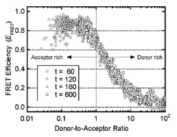
of two dye lipids are constant, 1 mol %, at any χ. By analyzing the data in Fig. 1B using these reasonable assumptions,
the quantitative determination of FRET efficiency depending on the donor-to-acceptor
ratio becomes possible as shown in Fig. 2. A large number of data follow
a unique curve, which supports the reliability of our experiments.
[1] K. Furukawa, et al., Lab Chip 6 (2006) 1001.
[2] K. Furukawa, et al., Langmuir 24 (2008) 921.
 |
 |
|||||
|
|