Å@
1Materials Science Laboratory, 2University of Oxford
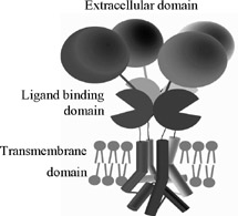
Å@Å@Ionotropic receptors (ligand-gated ion channel receptors) are important
membrane proteins for signaling in the neuronal networks of the central
nervous system. They are regulated by a ligand which binds to the extracellular
ligand binding domains. This binding gives an allosteric change in the
structure and opens the ion channel incorporated in the transmembrane domain
to allow cations flow into the cell (Fig. 1). Atomic force microscopy (AFM)
enables the nano-scale observation of proteins in a liquid environment,
offering a unique opportunity to observe functional biological molecule
such as single proteins under physiological conditions.
Å@Å@In this study, we have succeeded in observing the structure of single
purified and ionotropic receptor proteins in the solution using the AFM.
Receptor proteins were purified from over-expressed insect cells, and then
reconstituted into an artificial lipid bilayer by dialysis because the
receptors would function as in vivo when reconstituted into the lipid bilayer. We then imaged the reconstituted
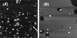
receptor proteins on a substrate in a buffer solution using AFM. Before
reconstitution, receptors were observed at the edge of small lipid patch
on mica (Fig.2 (A)), while after the reconstitution, receptor proteins
settled in the large lipid domain on mica (Fig. 2(B)) which provides for
the structural observation of the single receptor. Then by zooming in on
the single receptor protein, we could observe that it consisted of four
protrusions suggesting the four subunits of the receptor protein.
Å@Å@This study demonstrates that AFM can observe functioning single ionotropic
receptors, which suggests the possibility of determining a real-time conformational
change of a functioning membrane protein using AFM [1].
Å@Å@This research was supported in part by Bio-nanotechnology IRC in U.K.
and by the Strategic International Cooperative Program, Japan Science and
Technology Agency (JST).
[1] N. Kasai, et al., Neurosci. Res. 58 (2007) S193.
 |
 |
|||||
|
|