and Keiichi Torimitsu
Materials Science Laboratory, *Suzuka Nat. Coll. Tech.
To fabricate a nano-bio device that functions with membrane proteins, we are trying to form micron-scale wells covered with a lipid bilayer on a Si substrate [1]. The membrane proteins in the suspended membrane will be able to function as well as membrane proteins in vivo. In this study, we used giant unilamellar vesicles (GUVs) several tens of microns in diameter to form a lipid bilayer suspended over the wells.
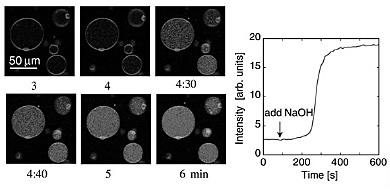
GUVs have been studied as a simplified cell membrane model. Figure 1 shows a functional analysis of ion channels (gramicidin A) arranged into a GUV lipid membrane based on an analysis of changes in fluorescent intensity. The GUVs’ internal environment includes the fluorescent probe (pyranine), which changes its emission intensity with pH. When the environment outside the GUVs is changed (pH is increased), the cations pass through the ion channels and cancel out the concentration gradient between the inside and outside of the GUVs. The increase in the pH of the GUVs with time is clearly shown.
Then, the GUVs were ruptured over the wells which were fabricated on the
Si substrate using photolithography. The aperture of the wells has an overhang
shape that was formed by selectively etching the Si substrate below the
SiO2 layer. The wells were loaded with fluorescent probes and sealed with a
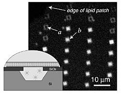
lipid bilayer. Excess probes were rinsed out. Figure 2 shows that the fluorescent
probes are trapped in the wells (b), although several holes around the patch are unfilled (a). The overhang shape improves the probability of the confinement of the
fluorescent probes in the wells because the suspended lipid bilayer is
prevented from falling into the wells. The fluorescence from the probes
confined in the wells remains unchanged for one hour or more. This indicates
that the probes remain stably in the wells without flowing out. When we
changed the pH outside the wells, we could analyze the function of the
ion channels in the suspended lipid bilayer, as in Fig. 1. We show that
micron-scale wells for the functional analysis of membrane proteins are
formed on the Si substrate.
The transport of ions through the ion channels will be analyzed by both the fluorescent intensity change and the ion current, which will be collected by an electrode embedded in the wells. This constitutes a major step toward the fabrication of nano-bio devices.
[1] Y. Shinozaki et al., Appl. Phys. Express 3 (2010) 027002.
 |
 |
|||||
|
|