Materials Science Laboratory,*University of Oxford
Membrane receptor proteins play the crucial role of transmitting signals in the central nervous system. They are regulated by chemicals called ligands which bind to the extracellular ligand binding domains. This binding either effects a change in the protein that forms a pore allowing movement of charged ions into the cell or modifies inside the cells chemically. As such, receptor proteins are small but highly selective devices. Most of the receptor proteins consist of multiple subunit proteins. The structure of the receptor proteins has been examined by mainly by X-ray crystallography and cryo-EM, however, these techniques do not allow the examination of functioning receptors. Atomic force microscopy (AFM) enables the nano-scale observation of proteins in a liquid environment, offering a unique opportunity to observe functional biological molecule such as single proteins under physiological conditions.
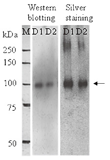
We have succeeded in observing the structure of single purified and ionotropic
receptor proteins in the solution using the AFM [1]. In this study, receptor
proteins were purified from over-expressed insect cells (Fig. 1), and then
reconstituted into an artificial lipid bilayer by dialysis because the
receptors would function as in vivo when reconstituted into the lipid bilayer. Receptor samples were settled
on to mica substrate and rinsed thoroughly. We then imaged the reconstituted
receptor proteins on a substrate in a buffer solution using AFM. We determined
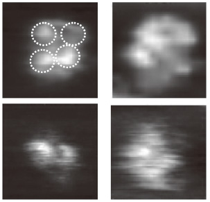
the orientation of the reconstituted receptor using antibody reaction,
and then we magnified and found four protrusions indicating tetrameric
structure of the protein (Fig. 2), which provides various different shapes
of the single receptor. This result first demonstrated that the structure
of the functioning receptor proteins seems to change mainly by heat fluctuation.
This research was supported in part by Bio-nanotechnology IRC in UK and
by KAKENHI .
[1] N. Kasai et al., BBA Gen. Subj. 1800 (2010) 655.
 |
 |
|||||
|
|