Si and Au Nanopillar Arrays as Scaffolds for Neuronal Growth
Neuronal in vitro cultivation has been widely used with the aim of elucidating neuronal signaling mechanisms and for applications in the neuro-engineering field. Our group has cultivated neurons on various substrates to create interfacial devices for neuronal guidance and thus realize artificial synapses. Neuronal guidance using nano-scale structures has also attracted attention thanks to recent advancements in nanotechnology. In this study, we examined neuronal guidance using nanopillar arrays made of amorphous silicon (a-Si) and gold (Au) as scaffolds for neuronal growth [1].
Nanopillars 100 and 500 nm in diameter were fabricated on quartz substrates using electron-beam lithography, and rat cortical neurons were cultivated on them for 7 days. The samples were then fixed and observed by using scanning electron microscopy and confocal laser scanning microscopy after the treatment.
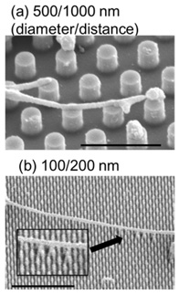
Neurons cause neurites to lengthen on an a-Si pillar as shown in Fig. 1. The neurites behaved differently in terms of width; they were wider on 500-nm-diameter pillars than on 100-nm-diameter pillars. This implies that the adhesion of neurites to pillars promotes skeletal protein expression, and thus neurite width is dependent on adhesion area size.
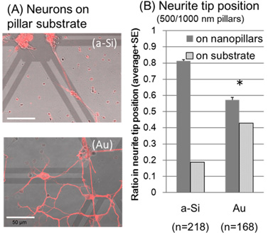
Then we examined neurons on different pillar materials as shown in Fig. 2. Neurons grew randomly on Au pillars while they became longer along with the patterns on a-Si pillars
[Fig. 2(A)]. A quantitative analysis demonstrated that there was a higher ratio of neurite tips on the a-Si pillars than on the Au pillars [Fig. 2(B)]. This low affinity of neurons for Au corresponds to neuronal cross-sections results obtained using FIB/SEM, which showed less attachment of soma to the Au substrate [2]. These results demonstrate the possibility of neuronal guidance using nanopillars made of appropriate materials.
This work was supported by JSPS/MEXT KAKENHI Grant Number 15H03541.
- [1] N. Kasai, R. Lu, R. Filip, T. Goto, A. Tanaka, and K. Sumitomo, Electrochemistry. 84, 296-298 (2016).
- [2] T. Goto, N. Kasai, R. Lu, R. Filip, and K. Sumitomo, J. Nanosci. Nanotechnol. 16, 3383 (2016).
|
|
| Fig. 1. Neurites grown on different patterned a-Si pillars. Scale: 2 μm. | Fig. 2. Neurons grown on a-Si or Au pillar substrate (A) and neurite tip position on different pillar materials (B). Scale: 50 μm. |