Electron Paramagnetic Resonance Using a SQUID Magnetometer Directly Coupled to an Electron Spin Ensemble
Magnetic resonance is a widely-used tool from analyzing material properties to medical applications, for instance with electron paramagnetic resonance (EPR) or nuclear magnetic resonance. However, conventional EPR spectrometers require a large number of spins (~1013) for the detection and do not have high spatial resolution (~0.1 mm). To expand the field of applications including quantum information processing, highly sensitive EPR spectrometers are necessary.
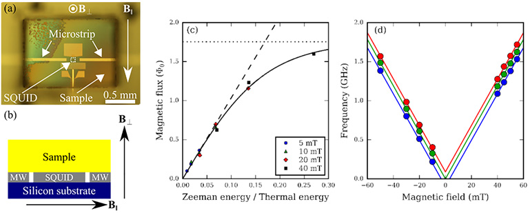
Here, we demonstrate the detection of electron spin polarization and EPR spectroscopy using a SQUID magnetometer directly coupled to an electron spin ensemble [1]. Our experimental setup is shown in Figs. 1(a) and (b) where the magnetization from the sample is detected by measuring the critical current of the SQUID. To begin, we measure the electron spin polarization as a function of the in-plane magnetic field and temperature. By increasing the in-plane magnetic field, we observe an increase of electron spin polarization ratio due to the reduction of the thermal fluctuations [Fig. 1(c)]. Figure 1 (d) shows the EPR spectrum of a type Ib diamond crystal containing nitrogen impurities. We confirm a linear increase of resonance frequency. Furthermore, ~93 MHz shifted peaks are observed due to hyperfine splitting of 14N.
The minimum detectable number of spins for our method is ~106. This number is much smaller than the conventional EPR spectrometer and can be improved for three orders of magnitude by replacing the SQUID with a superconducting flux qubit. Further we estimate the sensing volume at ~10-10 cm3 [~0.1 pL]. This is two order of magnitude smaller than the reported value in the literature [2].
This work was supported by the Commissioned Research of NICT, in part by an MEXT Grant-in-Aid for Scientific Research on Innovative Areas, and in part by KAKENHI grant.
- Present address: *Chinese Academy of Sciences
- [1] H. Toida et al., Appl. Phys. Lett. 108, 052601 (2016).
- [2] A. Bienfait et al., Nature Nanotech. 11, 253–257 (2016).
|
| Fig. 1. (a) Optical microscope image of the sample. (b) Cross sectional view of the experimental setup. (c) Magnetization as a function of the in-plane magnetic field and the temperature. (d) Results of EPR spectroscopy. |