
Research topics
I'm exploring the unique methods and physics for constructing systems and structures
at micron or submicron scales that are applicable for biology, microbiology, and tissue engineering.
My primary research interests lie at the intersection of micro-engineering, biology, and material science.
Especially, I'm focusing on the application of photo-lithography to soft materials to
manipulate individual cells and create their immediate extracellular environments.
This miniaturized tool and system have not oly the capability to transform the conventional study
on cellular physiology and behavior, but also the potential for developing cell-friendly interfaces
to measure cellular electrical signals toward medical diagnostics.
Because of their simplicity, low cost and use of MEMS process,
I will utilize a range of experimental methods and multidisciplinary knowledge
in order to develop new micro-/ nano-engineered biological analysis tools and implantable electrodes.
Manipulation of
adherent cells
Conductive
bio-interfaces
Transformable thin film
Microfluidic
cell manipulation
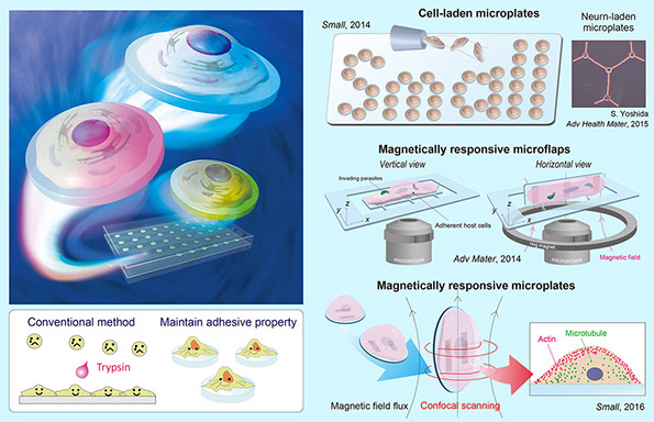
Manipulation of adherent cells
An approach for manipulating adherent cells is developed by integrating with an non-cytotoxic batch release. This strategy uses an array of releasable microfabricated mobile substrates, termed microplates, micro-pallets, and microflaps, formed from a biocompatible polymer, parylene. The parylene surfaces are modified with not only ECM to enhance cell attachment and growth, but also various functional materials. This system allows the inspection of selected single adherent cells while maintaining their adhesive properties, which broadens the examination of a variety of attributes, such as cell shape and cytoskeletal properties. For cell manipulation, the cell-laden films possess multiple functions: (i) magnetic response; (ii) electrical conductivity; and (iii) transformability. All of these cell interfaces are potentially applicable for not only the fundamental experimental tools for cell biology, but also the kits for drug-screening assay and platform for regenerative medicine. For example, magnetically responsive films can incline the loaded adherent cells in the desired orientation. Precisely controlled inclination of loaded cells by the applied magnetic field allows us to observe cell membrane boundaries from multiple angles, and obtain clear focused images of a type of parasite (Toxoplasma gondii) invading into their host cells with high magnification.
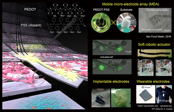
Conductive biointerface
As the interest in healthcare has grown in recent years, the technologies to monitor vital data have been attracting a lot of attention with a view to disease diagnosis and prevention as well as lifestyle improvement. I'm working on the research and development of conductive composite materials by coating or mixing conductive polymer with biocompatible substrates such as silk bundles and nonfibers. These composite materials are used as the electrodes for measuring biological signals from, for example, the cells, skins, or the heart, with high biocompatibility and sensitivity. I focus on utilizing conductive polymer (PEDOT:PSS) and graphene as non-cytotoxic and flexible electrodes. The microfabricated conductive cell-interfaces are applicable for not only the mobile multi-electrode arrays to record or apply neural signals, but also ex vivo regenerative medicine of artificial tissues. In addition, the tissue-interfaces integrated with flexible and stretchable electrode nanofiber (hitoer®) are applicable for continuously monitoring heart rate and for measuring ECG, EMG, and EEG waveforms only by wearing them. The vital data obtained from the patients, workers, and sport athletes during long-term use are very important because they become significant big-data to be analyzed with a view to disease diagnosis and prevention as well as lifestyle improvement.
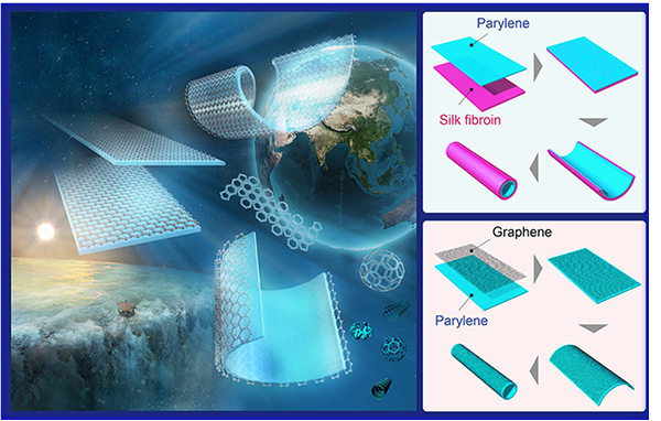
Transformable thin-film
My research goals are directed toward understanding molecular self-assembly into threedimensional (3D) shapes. My current research is focusing on micro-engineered 3D soft matter interfaces with biological samples like cells and tissues. This work is a fundamental step toward the bottom-up 3D fabrication, where small-scale 3D electrodes are built hierarchically by understanding and engineering thin film self-folding mechanism at the micro- and nano-scales. Since it draws on a diverse set of disciplines including soft materials, biology, and engineering, my approach of this research topic will have great impact on three fields including material science, biomedical science, and information technology.
Multi-layered thin films transform into various 3D geometries spontaneously due to heterogeneous mechanical properties. I'm developing transformable all-polymer films, termed micro-rolls, for 3D biointerfaces. These 3D interfaces are composed of various types of soft biocompatible materials including synthesized polymers, proteins, and 2D materials in order to add the functions to micro-rolls. Owing to their high biocompatibility and structural stability, they are potentially applicable for the reconstruction and assembly of functional tissues and implantable tissue grafts.
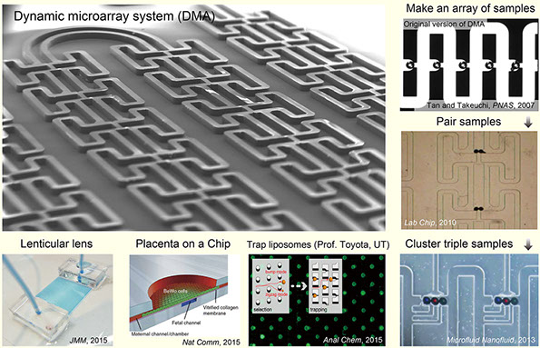
Microfluidic cell manipulation
As a fluidic interface with cells, I have developed the microfluidic system in which bio-molecules, chemicals, and single cells are immobilized toward biomedical screening process and high-throughput assay applications. For example, the dynamic microarray systems can trap, pair, array, and release micro-scale biological samples in the hydrodynamic sample traps in order to monitor them and observe their interactions. Various samples including single cells, spheroids, liposomes, microbeads can be manipulated in this microchannels in a high throughput manner. For instance, by mimicking in vivo, the pair two different types of cells are trapped in trapping sites to analyze cell-cell interactions. I can also apply microfulidic channels for organ-on-a-chip platform to mimick physiological systems of biological organssuch as brain, skins and placentas. This platform allows us to reconstitute 3D placenta tissues in vitro to recapitulate the physiology in an organ-specific context, and provides us a new finding that a fluid shear stress serves as a trigger for microvilli formation in placental cells. These microfluidic technologies will be alternatives to conventional cell culture models and animal testing for pharmaceutical and toxicology applications.

Updated on March 1st, 2020