Materials Science Laboratory
Cell membranes play an important role in the activity of bio-molecules,
for example, membrane proteins. A lipid bilayer, which is a basic component
of a cell membrane, can be fabricated artificially on a hydrophilic surface
by spontaneous growing characteristic (self-spreading). Such membranes
remain the fluidity based on lateral diffusion. Using these characteristics,
in this study, we investigated the dynamics of a lipid bilayer on a nano-patterned
surface and the molecular transport using the lipid bilayer as a molecule
carrier [1].
L-α-Phosphatidylcholine (extracted from egg yolk) containing 5 mol% of
dye-conjugated lipid was prepared. Dyes used were Texas Red, fluorescein
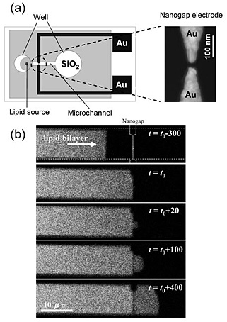
and NBD with different molecular size. Nanogap (10〜200 nm) using Au was
fabricated on an SiO2 surface. Microchannel that had wells at both ends was fabricated on this
nanogap structure using a photoresist [Fig. 1(a)]. A small amount of a
lipid source was adhered inside the well. The self-spreading of a single
lipid bilayer was initiated by immersing the device in a buffer solution.
Fluorescence from the lipid bilayer was observed by a confocal laser scanning
microscope.
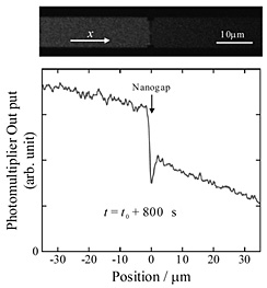
Figure 1(b) shows a time evolution of a self-spreading lipid bilayer before and after passage through a nanogap. The single lipid bilayer developed along a microchannel and successfully passed through the nanogap without development on photoresist and gold pattern. Figure 2 shows a fluorescence image and corresponding fluorescence intensity profile obtained after a sufficient time interval. This result clearly shows that the fluorescence intensity decreases discontinuously in the vicinity of the nanogap. This behavior strongly depends on the size of the nanogap and dye moiety. The dye molecules experience interference when they pass through the nanogap although the nanogap size is much larger than that of the dye moiety (at most 3 nm).
[1] Y. Kashimura et al., Jpn. J. Appl. Phys. 47 (2008) 3248.
 |
|
||
 |
|||
|