Materials Science Laboratory
Microarrays of biological molecules such as DNA chips are widely used
for high-throughput bioanalysis. When developing new microarrays, it is
essential to improve the technology for fixing biomolecules without losing
their biological functionality. A microarray consisting of an artificial
cell membrane supported on a solid surface is expected to provide an important
platform for the purpose. We have developed a new technique for fabricating
an artificial cell membrane microarray and demonstrated a biosensing application
of the fabricated microarray.
We used the self-spreading phenomenon, which is a self-organizing process
that forms a cell membrane at a solid-liquid interface, to fabricate the
artificial cell membrane microarray. Our method is unique in that the position
of the self-spreading is controlled with a hydrophilic/hydrophobic pattern
[1]. According to the designed pattern, a self-spreading membrane starting
from a macroscopic area was guided to the microarray structure without
mixing with membranes in the different areas. We succeeded in fabricating
10-µm-width parallel lines, each filled with an artificial cell membrane
with a unique composition, at 5-µm intervals (color frontispiece).
Structures obtained with our new technique are in principle more than 100
times more highly integrated than previously reported structures that employ
the vesicle fusion technique on patterned surfaces [2].
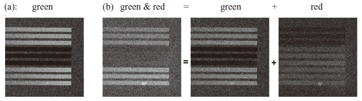
To demonstrate the validity of the microarray for biosensing applications, we fabricated a composition microarray, part of which contained biotin-conjugated lipid. Figure 1 shows fluorescence images of the array that were recorded (a) before and (b) after it was immersed in Texas Red conjugated streptavidin solution. Red fluorescence, which originates from Texas Red, was observed only in the lines containing biotin-conjugated lipids after 90 min, as shown in Fig. 1(b). This is due to the specific binding between biotin and streptavidin. Since the red fluorescence from the lines without biotin is limited, the microarray is valuable for biosensing applications.
This work was supported by KAKENHI.
[1] K. Furukawa et al., Lab Chip 6 (2006) 1001.
[2] K. Furukawa and T. Aiba, Langmuir 27 (2011) 7341.
 |
||
|