


Reconstitution of a functional neural network using a nanoscale thin-film-based 3D template
- new biological interface for regenerative medicine technology -
NTT Basic Research Laboratories demonstrated a method to assemble polymeric and cell-friendly thin films into three-dimensional structures. Since the materials of these films are not harmful to living organisms, neurons could be encapsulated inside them for a long period of time, during which they grew axons or neurites along the 3D structure and expressed their intrinsic functions. This achievement promises new biological cell transplantation interfaces for regenerative medicine and monitoring of living tissue.
The motivation behind this advance is that since biological samples are fragile, it is technically difficult to precisely to form them into a desired shape at the desired position. A sub-millimeter-scale artificial structure composed of biocompatible, flexible, and durable materials would allow such samples to be formed or grown in various shapes without damaging them.
Researchers at NTT have been studying a carbon-based nanomaterial called graphene1 for many years. They discovered that a graphene-laden polymeric thin film spontaneously assembles into a three-dimensional structure when it is released from the substrate supporting it. Poly-para xylylene (parylene2) was used as the material of the polymeric thin film, because it tightly sticks to graphene. The assembled 3D structure was used as a cell scaffold on which hippocampal neurons were successfully cultured and grown into functional fiber-shaped neuronal tissue. The fiber-shaped neuronal tissues were found to exhibit cell-cell interactions3 by forming a network in which they were connected to external neurons through minute holes in the self-folded tubular film.
Graphene and polymer thin films are non-toxic and have good affinity with living organisms. It is possible to assemble these films into various 3D structures by changing their shape and thickness. Moreover, owing to the excellent conductivity of graphene, the technology developed in this study can be applied to new biological interface devices such as the flexible electrodes for monitoring the periphery of transplanted cells in regenerative medicine, repairing damaged tissues, and assembling 3D artificial tissues for drug-discovery screening.
The Bio-Medical Informatics Research Center (BMC) at NTT was established in July 2019. The center’s mission is to apply the basic and applied research knowledge of materials science to the field of biofunctional materials and create ICT data-driven medicine and behavioral information analysis technology by cooperating with NTT Research Inc., the Medical & Health Informatics Laboratories, NTT operating companies, and hospitals owned by NTT Group.
=> Press Release
=> Molecular and Bio Science Research Group
Background of the research
Recent advances in medical technology and information processing technology have brought prospects for biological interface (bio-interface) that allow direct access to and monitoring of living biological tissue. Since the bio-interfaces should be durable, soft, and, compatible to the tissues, the search is on all over the world for new materials that can meet these requirements.
Graphene, which is essentially a single layer of carbon atoms, is one of the most promising two-dimensional (2D) functional materials. This extremely thin sheet-like material has high transparency, strength, chemical resistance, heat resistance, flexibility, and biocompatibility. Thanks to these characteristics, it is being examined as a potential alternative to using silicon and rare metals in transistors, batteries, and sensors. However, because graphene is an extremely thin 2D sheet, it has proven difficult to assemble and refine it into a three-dimensional (3D) structure with sufficient definition to be practically used in the above devices.
A method for processing and bending graphene freely and easily has not been established, and there are still technical problems that cause fracture and exfoliation. If graphene can be formed into 3D structures, it could be used as an interface that fits the surface of biological samples. As such, it could be used to make in vivo implantable electrodes and microscale cell-culture substrates. Therefore, graphene is potentially useful in regenerative medicine and for making medical/therapeutic devices.
Research results
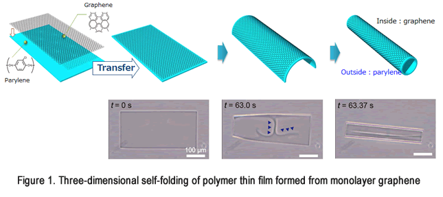
We developed an easy method to assemble graphene into a fine three-dimensional structure, by transferring it from a glass substrate to a parylene polymer thin film. The transferred graphene adheres strongly to the parylene film and spontaneously deforms it into a 3D structure when it is released from the substrate due to the difference in the elastic moduli of the materials (this phenomenon is called self-folding). This method enables us to finely bend graphene with a curvature radius of 4 µm at a minimum by changing the thickness of the polymer thin film (Figure 1, Movie 1). In addition, all of the materials used in this technology are cell-friendly and highly flexible. We used self-folding 3D graphene formed into a cylinder as an interface with biological samples (cells). This self-folding graphene scaffold encapsulated the cells (neurons in this case), and the cells were stably cultured inside its hollow center it for a long time. In particular, we succeeded in artificially growing a minute and tissue-like nerve structure along the scaffold. We formed a network between it and other neurons outside the scaffold and demonstrated cell-cell interactions both inside and outside the structure.
Technical point: Self-assembly of 3D graphene structure that is compatible with cells
- Three-dimensional structure of graphene using polymer thin film
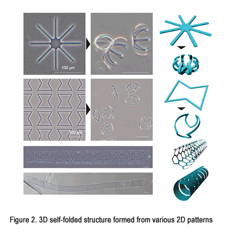
The final self-folded three-dimensional shape depends on the original two-dimensional micropattern and the thicknesses of the thin film. For example, a finer structure can be produced by thinning the thin film; thin films with rectangular, parallelogram, and radial micropatterns can be spontaneously assembled into cylinders, coils, and spheres, respectively (Figure 2). In addition, it is possible to increase the permeability of the structure to molecules from the outside by making tiny holes in a rectangular two-dimensional pattern. These tiny holes allow nutrients and oxygen to be supplied to the encapsulated cells for a long period and thereby provide a means of stable culture. - Application of 3D graphene structure to interfaces with cells
Since the materials constituting the 3D graphene structures are not cytotoxic, it is possible to encapsulate cells inside them (Figure 3, Movie 2). In addition, their high transparency enables us to observe and analyze the cells using various microscopes. The encapsulated cells can be from cell lines or primary cells that are isolated from other living tissues. In addition, by controlling the density of the encapsulated cells and the shape of the three-dimensional structure, we can produce cell aggregates with any shape after culturing for about a week. For example, when the hippocampal neurons were cultured in the cylindrical structure, the cells aggregated and formed fiber-like artificial nerve tissue. The excellent robustness and stiffness of the graphene and parylene led to a stable and long-term cell culture without any disruption of the cells. - Long-term culture of neurons and creation of neural network
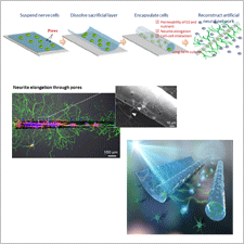
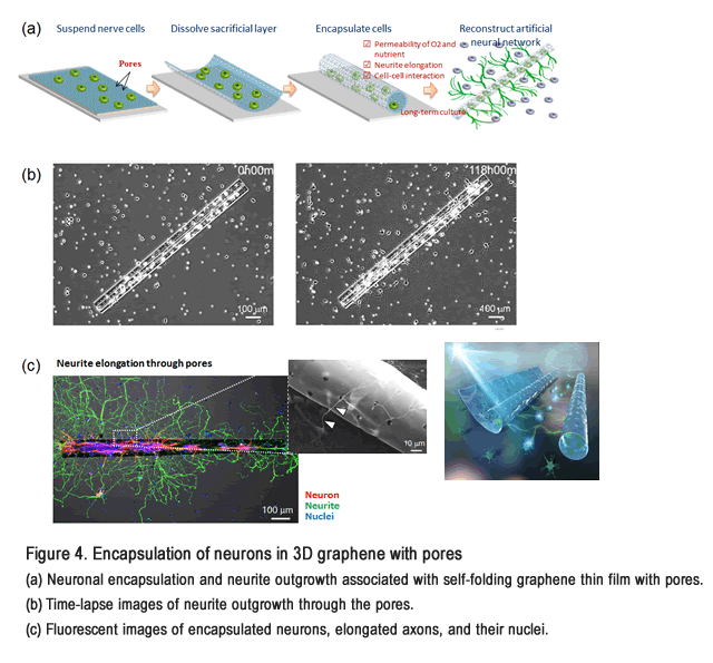
Nutrients and oxygen in a culture solution can easily flow through a 3D graphene structure with many pores. This enables a more stable and longer culture compared with the one in which the graphene did not have pores (Figure 4, Movie 3). Furthermore, we observed that the neurons inside the structure never passed through the pores, but extended their neurites through them (Figure 4). We could thus connect the neurons inside the structure to ones outside it via these extended neurites. The resulting neuronal network exhibited synchronization of spontaneous neural activities. This result indicates that by extending axons through the pores, functional synaptic connections can be formed and neurons in the scaffold can be functionally integrated into surrounding neuronal networks (Video 4).
Future prospects
We developed a technology to spontaneously form graphene into any desired shape and produce transparent biocompatible 3D structures. We experimentally encapsulated and stably cultured cells inside a self-folded 3D structure. Our method can be used to make scaffold structures for regenerative medicine and examine the behaviors of single cells; it can also be used to make new bio-interfaces, for example, in vivo implantable electrodes that utilize the high conductivity of graphene.
Publication details
Authors: T. F. Teshima, C. S. Henderson, M. Takamura, Y. Ogawa, S. Wang, Y. Kashimura, S. Sasaki, T. Goto, H. Nakashima, and Y. Ueno
Title: Self-Folded Three-Dimensional Graphene with a Tunable Shape and Conductivity
Journal title: Nano Letters
Publication date: 9th, January, 2019
Authors: K. Sakai, T. F. Teshima, H. Nakashima, and Y. Ueno
Title: Graphene-based neuron encapsulation with controlled axonal outgrowth
Journal title: Nanoscale
Publication date: 28th, July, 2019
Description of the experiments
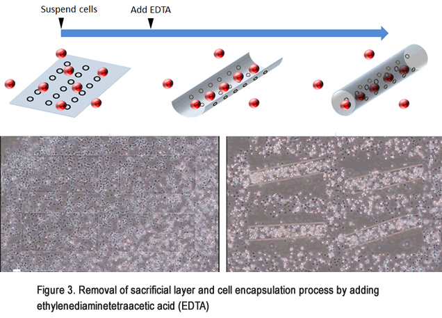
Two types of materials were used to make the bi-layer thin film: monolayer graphene sheets and polymeric parylene with thicknesses ranging from 50 nm to 500 nm. In addition, a calcium alginate hydrogel layer was placed underneath the graphene/parylene bilayer4 as a sacrificial layer. We could control the film thickness of both the graphene and parylene and process them into any two-dimensional pattern by using lithography technology.
Calcium alginate gel can be dissolved instantly by adding chelating agents such as ethylenediaminetetraacetic acid (EDTA5) or sodium citrate solution. This non-cytotoxic dissolution of a sacrificial layer allows the release of the bilayer membrane from the substrate. In the experiment, the calcium alginate sacrificial layer gradually dissolved from the outer periphery of the thin film, which led to the thin films deforming into a 3D shape. In particular, rectangular films were deformed into a cylindrical structure such that the graphene was on the outside surface. This transformation of graphene-laden parylene films encapsulated the cells without damaging them.
Glossary
- *1 ... graphene
- Graphene is a sheet-like material with the thickness of a single atom in which carbon atoms are covalently bonded and form a hexagonal lattice honeycomb network. Graphene can be easily obtained by peeling it from the graphite used for pencil leads. Its high electron mobility, transparency, chemical/thermal stability, and superior flexibility compared with conventional semiconductors have been motivations to apply it to transistors, sensors, and flexible electronic circuits.
- *2 ... poly(p-xylylene) (parylene)
- Parylene is a polymer with a linear crystalline structure that is a good insulator and acts as a strong barrier against moisture and chemical substances. In addition, it has excellent heat resistance and ultraviolet resistance and is highly biocompatible. It is used as a coating agent in a wide range of industrial fields including medical care.
- *3 ... cell-cell interaction
- The cells of multicellular organisms interact with each other to form various tissues and maintain homeostasis. These cell-cell interactions are broadly classified into direct cell-cell interactions, signaling molecule-mediated interactions, and neuron-to-neuron electrical interactions. The intercellular interactions of cells in the 3D graphene structure are exchanges of electrical signals between nerves.
- *4 ... calcium alginate gel
- Alginate acid is a polysaccharide that can be extracted from seaweed such as kelp and sea mustard. It consists of two types of monosaccharide (mannuronic acid and guluronic acid) joined in a straight chain. Calcium alginate gel is formed when these saccharides are cross-linked by calcium ions, and it swells up with the addition of water. This allowed the thin film in the experiment to be liberated from the glass substrate without harming any of the cells.
- *5 ... ethylenediaminetetraacetic acid (EDTA)
- Ethylenediaminetetraacetic acid is a chelating agent commonly abbreviated as EDTA. It binds strongly with divalent ions such as calcium to form a complex. It is widely used in industry for the removal of heavy metals from fiber pulp, medicines, and foods, and it is used in biology to remove calcium and deactivate enzymes. In this study, it was used to remove calcium ions from the calcium alginate layer4, thereby dissolving the layer and releasing the thin film.
Video Links
(Video 1) Self-folding of graphene / parylene bilayer
(Video 2) Damage-free cell encapsulation in 3D graphene
(Video 3) Pore-mediated elongation of neurites
(Video 4) Synchronization of nerve cell activity


